Primary Liver Tumours
Hepatocellular Carcinoma (HCC)
Hepatocellular carcinoma is cancer of the liver, which arises from liver cells (hepatocytes).
Hepatocellular carcinoma Q & A
Cancer of the liver usually occurs in people with liver disease, such as liver cirrhosis, which is commonly caused by hepatitis virus infection, heavy drinking or having a fatty liver. It can also arise from a benign tumour of the liver (liver cell adenoma). Rarely, it can occur in an otherwise normal liver.
The symptoms are often vague. They may include weight loss, abdominal pain or discomfort, feeling unwell and lethargic, and having nausea and distension of the abdomen.
Blood tests are usually performed, including testing for a substance called alpha-fetoprotein (AFP), which is raised in some people with cancer of the liver.
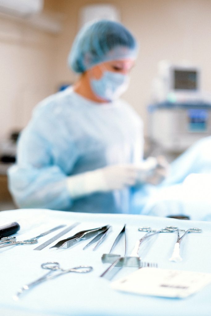
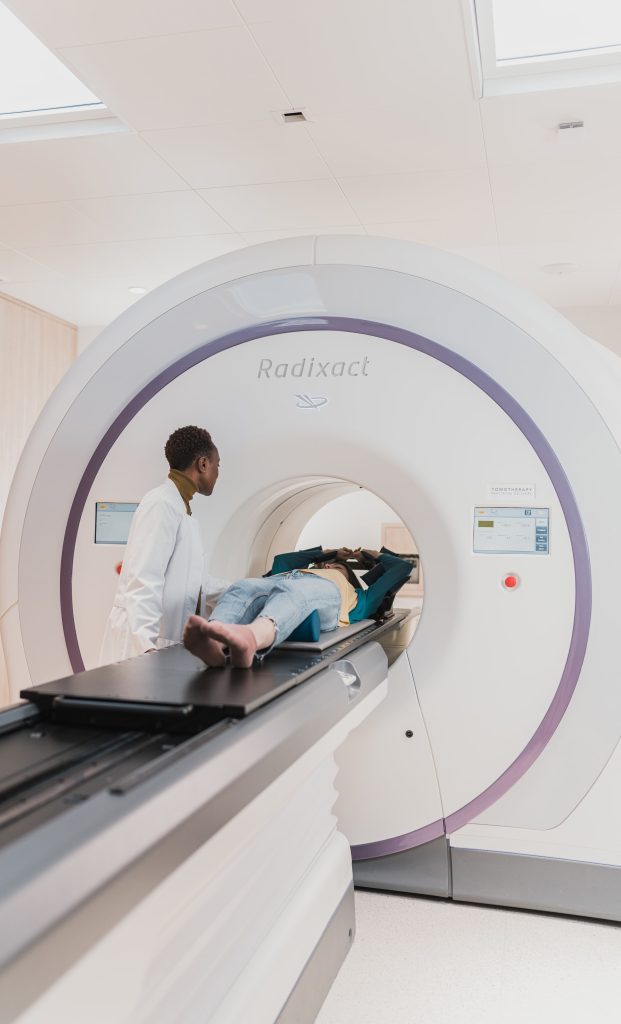
Scans including ultrasound, triple phase computerised tomography (CT) and magnetic resonance imaging (MRI) of the liver are usually performed. In certain cases a biopsy may be needed.
The treatment depends on several factors such as the size, location and number of tumour(s), the state of the liver (degree of cirrhosis and function) and fitness of the patient.
Liver transplantation is offered to patients with liver cirrhosis. The patient needs to meet certain criteria for transplantation to be considered.
Surgical resection to remove the tumour from the liver may be possible in patients who do not have cirrhosis, or patients with cirrhosis that have preserved liver function but are not suitable for liver transplantation.
Patients not suitable or ready for either of the above treatments may be offered treatments that may improve symptoms and slow the cancer. Trans-arterial chemo-embolisation (TACE) delivers chemotherapy directly to the tumour and blocks the blood supply. Radio-frequency ablation (RFA) generates heat which destroys tumour tissue.
Chemotherapy with a drug called sorafenib may be offered to patients who have advanced disease not suitable for the treatment options above.
Secondary Liver Tumours Q & A
Yes. Surgery to remove tumours from the liver (hepatectomy) is routinely performed for secondaries arising from cancer of the colon or rectum. Secondaries from other tumour types may also be amenable to surgery. This includes:
Neuro-endocrine tumours
Gastro-intestinal stromal tumours (GIST)
Renal cell (kidney) cancer
Surgery is also offered in selected patients with secondaries from:
Breast cancer
Melanoma
Nasopharyngeal cancers
Thyroid cancer
Bowel cancer is the commonest gastro-intestinal cancer in the UK. Up to half of the patients with bowel cancer develop secondary spread to the liver at some stage of their illness. A significant proportion of patients with liver secondaries will be found suitable for surgery after careful assessment by liver specialists (a surgeon, radiologist and oncologist). Others who may not be suitable for liver surgery initially may become eligible after a period of treatment with chemotherapy.
Surgery to remove liver secondaries from colorectal cancer can result in up to half of the patients living for 5 years or more, and a fifth of the patients living for 10 years or more. This surgery provides patients a realistic chance of being cured of their disease. Although chemotherapy is effective alongside surgery in this type of cancer, it does not, on its own, provide a cure.
Liver secondaries are usually picked up on an ultrasound or computerised tomography (CT) scan. Patients, whose primary cancer has not spread, initially undergo surveillance with regular scans to pick up any secondaries that may develop.
If these are suspected further tests are offered, including a magnetic resonance imaging (MRI) scan of the liver as well as a positron emission tomography (PET) scan.
The liver is the only organ that has the capacity to regenerate. It is possible to remove about two thirds of the liver volume safely.
In certain cases it is possible to induce growth of a future liver remnant by blocking the portal vein to the opposite half of the liver (i.e. the half containing tumours).
This technique, called portal venous embolisation, has made it possible to perform liver surgery for patients who were initially thought to be inoperable.
Radiofrequency ablation (RFA) can be used to destroy tumour deposit using heat in suitable circumstances.
Selective Intra-arterial Radiation Therapy (SIRT) involves the infusion of radioactive microspheres into the artery supplying the liver. These microspheres lodge in the tumours and target tumour cells.
Benign Liver Lesions
These are non-cancerous abnormalities in the liver. They include haemangiomas, focal nodular hyperplasia and adenomas.
Benign Liver Lesions Q & A
A liver haemangioma is made up of abnormal blood vessels. It is the commonest benign lesion of the liver.
Haemangiomas do not usually cause symptoms and are usually found by chance on scans that are performed for other reasons. Surgery is not indicated in most cases.
Focal nodular hyperplasia (FNH) is the second most common benign tumour of the liver.
These tumours occur more commonly in young women. Again, they are usually discovered by chance on scans performed for another purpose. They do not usually cause symptoms, so surgery is not usually needed unless there is uncertainty about the diagnosis. Your doctor may keep you under observation.
Liver (hepatocellular) adenomas are not as common as haemangiomas or focal nodular hyperplasia. They most commonly occur in women.
Although most do not cause symptoms, some can rupture and bleed. Surgery is usually recommended because there is a risk of developing liver cancer (hepatocellular carcinoma) from an adenoma and because of the risk of rupture and bleeding.
Liver Cysts Q & A
A liver cyst is a fluid collection which is surrounded by liver tissue. The liver tissue may be very thin in some areas.
Liver cysts affect approximately 5 in every 100 people.
Most liver cysts are called simple cysts. These are mostly single but multiple cysts can exist.
Some patients have a rare condition called polycystic liver disease. In this condition the liver contains a large number of cysts.
Rarely cystic tumours can occur in the liver. When these are benign, they are called cystadenomas. They have the potential to transform into cancer and are then called cystadenocarcinoma.
Most liver cysts do not cause symptoms. However, some can grow large enough to cause fullness in the abdomen and discomfort or pain.
Liver cysts are usually diagnosed on ultrasound scans.
A computerised tomography (CT) or magnetic resonance imaging (MRI) scan are performed if there is uncertainty about the nature of the liver cyst or to plan surgery.
Most simple liver cysts do not require treatment. Keyhole surgery to remove part of the cyst wall (fenestration) is performed when simple cysts cause significant symptoms. This is usually an effective and durable treatment.
Polycystic liver disease (multiple cysts that grow throughout the liver) may, uncommonly, cause such severe symptoms that liver transplantation is recommended.
Cystic tumours, whether cancerous or not, require a liver resection to remove the entire cyst. This is a more complex operation compared to fenestration of a simple cyst.